Public Lectures, Podcasts, Interviews, and Workshops
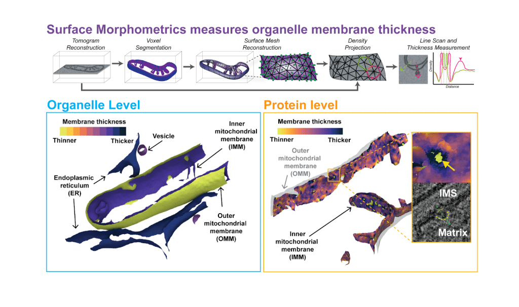
2026 Back to the density: using Surface Morphometrics to measure organelle membrane thickness in cryo-electron tomography
Michaela Medina presents our paper Surface Morphometrics reveals local membrane thickness variation in organellar subcompartments in a FocalPlane post!
2026 Structure Club Podcast
Danielle Grotjahn and Benjamin Barad present their paper, Surface Morphometrics reveals local membrane thickness variation in organellar subcompartments
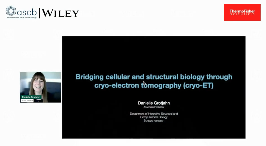
2026 ASCB/Wiley Webinar: Bridging cellular and structural biology through cryo-electron tomography
Cellular cryo-electron tomography (cryo-ET) holds the grand promise of integrating cellular architecture with molecular structure. Yet, for many cell biologists, this method can seem technically daunting or limited in scope compared to other imaging approaches. In this webinar, Prof. Grotjahn will discuss how cryo-ET is redefining the study of cellular organization by providing direct views of how macromolecules assemble and function in their native environment. Prof. Grotjahn will showcase her lab’s software, Surface Morphometrics, which converts three-dimensional reconstructions into quantitative models linking molecular structure to cellular mechanism. Prof. Grotjahn will highlight where cryo-ET excels, the challenges that remain, and how it fits within the growing landscape of other nanoscale imaging modalities. Explore how these advances are transforming the familiar cartoon models of cells into structural maps of molecular architecture.
2026 Evolving with Septins: Interview with Michelle Momany  .
.
2025 Cytoskeleton Spotlight: Michaela Horger  .
.
2025 Interviews with Authors, Jan-Hannes Schaefer
Single-particle cryo-electron microscopy (cryo-EM) has become an essential tool in structural biology. However, automating repetitive tasks remains an ongoing challenge in cryo-EM data-set processing. Here, we present a platform-independent convolutional neural network (CNN) tool for assessing the quality of 2D averages to enable the automatic selection of suitable particles for high-resolution reconstructions, termed CryoSift. We integrate CryoSift into a fully automated processing pipeline using the existing cryosparc-tools library. Our integrated and customizable 2D assessment workflow enables high-throughput processing that accommodates experienced to novice cryo-EM users.
2024 Front Row Lecture
The mitochondria are well known for being cellular “powerhouses,” given their important role in energy generation. Yet, emerging research is now suggesting these organelles also play a key role as the stress-sensors for the cell. In this free Front Row lecture, Scripps Research assistant professor Danielle Grotjahn explored how mitochondria change shape in response to different genetic and environmental stressors. By harnessing cutting-edge imaging technologies to examine mitochondria in these never-before-seen-ways, Grotjahn is revealing how these organelles can predict overall cellular health and even disease, including neurodegenerative disorders and cancer.
2023 Decoding the mitochondria with Dr. Danielle Grotjahn
Dr. Danielle Grotjahn, Assistant Professor at Scripps Research Institute, discusses how cryo-electron tomography is a powerful tool that is advancing our understanding of the mitochondria and ultimately human health.
2023 Science Changing Life Podcast
Are the mitochondria truly the powerhouses of the cell? In this episode, assistant professor Danielle Grotjahn shares why she thinks “the stress sensors of the cell” may be a more appropriate name for this cellular organelle–and more. Dr. Grotjahn works in the Department of Integrative Structural and Computational Biology at Scripps Research, where her lab is answering how mitochondrial networks change shape in response to genetic, pharmacological or environmental stress. Listen as we talk about the links between mitochondrial dysfunction and disease, cell death, and the cutting-edge imaging technologies that are enabling Grotjahn and her team to peer into the mysteries of the mitochondria.
2019 Scripps Research blog Part 1 and Part 2 A conversation with: Danielle Grotjahn, PhD, Scripps Research Fellow
![]()
2018 Radiobio interviews Dr. Danielle Grotjahn
We often imagine a cell as a large balloon filled with jelly, but really it is more like a large city. Packages need to go from one place to the other in an organized fashion as to not disrupt other processes. For example, when we need an item, we go to the store or click away on retail websites, but how do these items find their way to the retail place or our house? There are vehicles on roads and highways that are utilized for distribution. Much like the infrastructure that we use everyday to move cargo around our cities, the cell has its own system to deliver goods from one place to another. What are the 18 wheelers of the cell, how do they move such important packages, and how do they know where to go? Cytoplasmic dynein is a protein complex that transports molecular cargo along and plays a key role in the intracellular trafficking network. Dr. Danielle Grotjahn utilizes specialized imaging techniques to study these structures and the function of motor proteins.